Article, Cardiology

T-wave inversion: cardiac memory or myocardial ischemia?

Case Report

T-wave inversion: cardiac memory or myocardial ischemia? Abstract

This article presents a case report of a 74-year-old man with T-wave inversion (TwI) in atrial fibrillation noted during routine pacemaker interrogation.

The patient was seen for routine pacemaker interrogation, at which time he was noted to have underlying atrial fibrillation. A 12-lead electrocardiogram of the atrial fibrillation revealed significant TwIs. He was subsequently worked up for myocardial ischemia and was found to have a moderate-sized, moderate-degree inferior wall myocardial Perfusion defect. He was subsequently referred for a cardiac catheterization.

The cardiac catheterization revealed nonobstructive coronary artery disease. The follow-up electrocardiogram revealed persistent but attenuated TwI.

The TwIs were attributed to cardiac memory, a common but infrequently recognized phenomenon of which many

clinical practitioners are unaware. Cardiac memory is due to the T wave tracking the preceding abnormal QRS complex and can be induced by right ventricular pacing or arrhythmias.

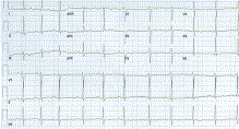
We present a case report of a 74-year-old man with new- onset T-wave inversion (TwI) noted during a routine clinic visit. His past medical history is significant for hypertension, Paroxysmal atrial fibrillation (Figs. 1 and 2), and sick sinus syndrome, for which a dual chamber pacemaker was implanted, and programmed (AAIR b-N DDDR mode) to minimize unnecessary right ventricular pacing (Fig. 3).

After routine pacemaker interrogation, a 12-lead electro- cardiogram (ECG) was obtained to document underlying atrial fibrillation because he had previously been in sinus rhythm on prior visits. The ECG documented atrial fibrilla- tion with narrow and upright QRS complexes but marked TwI in the inferior (II, III, and AVF) and anterior-lateral (V3-V6) leads (Fig. 4). The patient was asymptomatic and had no history of coronary artery disease, diabetes, or stroke. His

Fig. 1 Baseline normal sinus rhythm.

0735-6757/$ – see front matter.

Fig. 2 Baseline paroxysmal atrial fibrillation.

hypertension was well controlled on multiple agents: amlodipine 10 mg daily, hydrochlorothiazide 50 mg daily, losartan 100 mg daily, and metoprolol tartrate 25 mg BID.

An exercise/rest Tc99m Sestamibi stress test with gated SPECT was performed to rule out myocardial ischemia as a cause of the TwI. A moderate-size, moderate-degree

reversible inferior wall perfusion defect was noted, with a preserved left ventricular ejection fraction of 57%. Subse- quent cardiac catheterization revealed nonobstructive cor- onary artery disease. Attenuation of the TwI was documented 2 weeks later on a repeat 12-lead ECG. The final diagnosis was cardiac memory TwI (Fig. 5).

Fig. 3 Paced rhythm (right ventricular pacing, with occasional AV pacing).

Fig. 4 Postpacing electrocardiogram with TwIs.

Cardiac memory is a common but infrequently recognized phenomenon in which the T wave of the sinus or intrinsic rhythm tracks the QRS vector of the preceding abnormal

impulse [1]. It manifests on the ECG or vectorcardiogram as a TwI during sinus or any rhythm with normal ventricular activation and occurs after a period of altered myocardial

Fig. 5 Attenuation of TwI, 2 weeks after pacemaker clinic.

activation (such as after ventricular pacing, Left bundle branch block, ventricular tachycardia, and ablation of pre- excitation) [2]. The cellular mechanisms of cardiac memory remain unclear, but alterations in the transient outward current, Ito1, have been reported [3].

Cardiac memory may often be unrecognized, but it has potentially important clinical implications, such as alteration in the action of anti-arrhythmic drugs, with reduced efficacy or potential proarrhythmic effects [1]. Because cardiac memory can mimic TwI in myocardial ischemia, differ- entiation between these 2 events is important. Although there have been no established ECG criteria to distinguish between ischemic TwI and cardiac memory TwI, a recent study has proposed that the combination of a positive T wave in lead aVL, positive or isoelectric T wave in lead I, and maximal precordial TwI greater than TwI in lead III was 92% sensitive and 100% specific for cardiac memory, allowing discrimination from ischemic precordial TwI [4]. Using this criteria, the patient’s ECG met the criteria for cardiac memory.

Awareness of the cardiac memory phenomenon is important for health care providers, especially cardiologists and emergency department physicians, to facilitate appro- priate evaluation and management. Furthermore, although minimization of unnecessary right ventricular pacing is advocated to reduce the incidence of atrial fibrillation and heart failure, it may also help to reduce the occurrence of

cardiac memory, which can affect antiarrhythmic drug actions [1].

Carol Chen-Scarabelli MSN, APRN-BC

VA Ann Arbor/University of Michigan Healthcare System

Ann Arbor, MI, USA E-mail addresses: [email protected],

[email protected]

Tiziano M. Scarabelli MD, PhD

Wayne State University School of Medicine

Detroit, MI, USA

doi:10.1016/j.ajem.2008.10.037

References

  1. Patberg KW, Shvilkin A, Plotnikov AN, Chandra P, Josephson ME, Rosen MR. Cardiac memory: mechanisms and clinical implications. Heart Rhythm 2005;2:1376-82.
  2. Erdogan O. Spontaneous T wave inversion after ventricular pacing: what is the probable mechanism? Postgrad Med J 2006;82:e20-1.
  3. Yu H, McKinnon D, Dixon JE, Gao J, Wymore R, Cohen IS, et al. Transient outward current, Ito1, is altered in cardiac memory. Circulation 1999;99:1898-905.
  4. Shvilkin A, Ho KK, Rosen MR, Josephson ME. T-vector direction differentiates postpacing from ischemic T-wave inversion in precordial leads. Circulation 2005;111:969-74.

Leave a Reply

Your email address will not be published. Required fields are marked *