Measuring cardiac index with a focused cardiac ultrasound examination in the ED☆☆☆
Affiliations
- Department of Emergency Medicine, Loma Linda University, Loma Linda, CA 92354, USA
Affiliations
- Department of Emergency Medicine, Loma Linda University, Loma Linda, CA 92354, USA
Affiliations
- School of Medicine, Loma Linda University, Loma Linda, CA 92354, USA
Affiliations
- Department of Medicine, Division of Cardiology, Loma Linda University, Loma Linda, CA 92354, USA
Affiliations
- Department of Emergency Medicine, Loma Linda University, Loma Linda, CA 92354, USA
Affiliations
- Department of Emergency Medicine, Loma Linda University, Loma Linda, CA 92354, USA
Affiliations
- Department of Emergency Medicine, Loma Linda University, Loma Linda, CA 92354, USA
- Department of Medicine, Division of Pulmonary and Critical Care, Loma Linda University, Loma Linda, CA 92354, USA
Correspondence
- Corresponding author.

Affiliations
- Department of Emergency Medicine, Loma Linda University, Loma Linda, CA 92354, USA
- Department of Medicine, Division of Pulmonary and Critical Care, Loma Linda University, Loma Linda, CA 92354, USA
Correspondence
- Corresponding author.
 Article Info
To view the full text, please login as a subscribed user or purchase a subscription. Click here to view the full text on ScienceDirect.

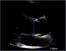
Fig. 1
Measurement of LVOT diameter at aortic valve cusps. This is a parasternal long-axis view on zoom mode with calipers measuring the LVOT diameter at 1.98 cm. This view is obtained by placing the probe at the fourth intercostal space lateral to the left sternum.
Fig. 2
Measurement of VTI using the apical 5-chamber view. This view is obtained with the apical 4-chamber view and then angulating the probe slightly toward the patient's head until the LVOT can be visualized. The pulsed-wave Doppler cursor is placed at the LVOT, and the doppler signal is traced (dotted lines) to calculate the VTI.
Fig. 3
Scattered plot for CI measured by ED physician compared with sonographer.
Fig. 4
Bland-Altman analysis for CI measured by ED physician compared with sonographer.
Abstract
Objectives
Noninvasive technology may assist the emergency department (ED) physician in determining the hemodynamic status in critically ill patients. The objective of our study was to show that ED physicians can accurately measure cardiac index (CI) by performing a bedside focused cardiac ultrasound examination.
Methods
A convenience sample of adult subjects were prospectively enrolled. Cardiac index, left ventricular outflow tract (LVOT) diameter, velocity time integral (VTI), stroke volume index, and heart rate were obtained by trained ED physicians and a certified cardiac sonographer. The primary outcome was percent of optimal LVOT diameter and VTI measurements as verified by an expert cardiologist.
Results
One hundred patients were enrolled, with obtainable CI measurements in 97 patients. Cardiac index, LVOT diameter, VTI, stroke volume index, and heart rate measurements by ED physician were 2.42 ± 0.70 L min−1 m−2, 2.07 ± 0.22 cm, 18.30 ± 3.71 cm, 32.34 ± 7.92 mL beat−1 m−2, and 75.32 ± 13.45 beats/min, respectively. Measurements of LVOT diameter by ED physicians and sonographer were optimal in 90.0% (95% confidence interval, 82.6%-94.5) and 91.3% (73.2%-97.6%) of patients, respectively. Optimal VTI measurements were obtained in 78.4% (69.2%-85.4%) and 78.3% (58.1%-90.3%) of patients, respectively. In 23 patients, the correlation (r) for CI between ED physician and sonographer was 0.82 (0.60-0.92), with bias and limits of agreement of −0.11 (−1.06 to 0.83) L min−1 m−2 and percent difference of 12.4% ± 10.1%.
Conclusions
Emergency department ED physicians can accurately measure CI using standard bedside ultrasound. A focused ultrasound cardiac examination to derive CI has potential use in the management of critical ill patients in the ED.
To access this article, please choose from the options below
Purchase access to this article
Claim Access
If you are a current subscriber with Society Membership or an Account Number, claim your access now.
Subscribe to this title
Purchase a subscription to gain access to this and all other articles in this journal.
Institutional Access
Visit ScienceDirect to see if you have access via your institution.
☆Funding source: This study was partially funded by the Emergency Medicine Research and Education Foundation.
☆☆Presentations: This study was presented at the American College of Emergency Physicians, Scientific Assembly, October 2011, San Francisco, CA.
Related Articles
Searching for related articles..


