Article, Urology

What do we miss without contrast in patients with flank pain?

Imprint logo

Case Report

Contents lists available at ScienceDirect

American Journal of Emergency Medicine

journal homepage: www. elsevier. com/ locate/ajem

American Journal of Emergency Medicine 34 (2016) 765.e3-765.e5

Journal logo

What do we miss without contrast in patients with flank pain?

Abstract

Flank pain is a common complaint in the emergency department, and the most frequent cause of flank pain is urolithiasis. Spontaneous renal artery dissection and renal Artery thrombosis are rare causes of abdominal pain which can result in renal parenchymal injury. They are mostly difficult to diagnose and treat in the emergency setting. The present report describes 2 patients admitted to the emergency department because of acute flank pain who were diagnosed with Renal infarction.

Flank pain is a common complaint in the emergency department (ED) which might be caused by a variety of extraurinary and urinary ab- normalities (eg, lower lobe pneumonia, zona zoster, urinary tract infec- tion, diverticulitis, cholecystitis, gastroenteritis). The most frequent cause is urolithiasis [1]. Spontaneous renal artery dissection and renal 
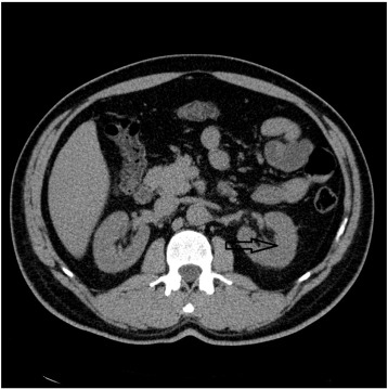
artery thrombosis are rare causes of abdominal pain which can result in renal parenchymal injury [2]. Spontaneous renal artery dissection and renal artery thrombosis are mostly difficult to diagnose and treat in the emergency setting. Ultrasound and Color Doppler examination is not sensitive enough to detect the dissection or renal infarction. A number of imaging modalities may be useful in diagnosis such as intravenous pyelogram, computed tomography (CT) scan, or magnetic resonance imaging. But the criterion standard is angiogram [3]. Some patients do well with conservative treatment, whereas others need emergent or Urgent surgery. Observation with anticoagulation, endoVascular procedures, open vascular surgery, and nephrectomy are proven as effective treatment modalities [4]. The present report describes 2 patients admitted to the ED because of acute flank pain who were diagnosed with renal infarction.

A 39-year-old man presented to the ED with a history of left-sided flank pain extending to the left iliac fossa region for about 3 days. The patient had no history of dysuria, hematuria, or fever. He stated that his pain had ensued suddenly while he was sleeping. There was no remarkable illness or medication in his medical history. He did not smoke or use alcohol or illicit drugs. Physical examination showed vital signs as follows: body temperature of 36.7?C, blood pressure of 170/93 mm Hg, heart rate of 84 beats per minute, respiratory rate of 17 beats per minute, and pulse oximetry of 99% on room air. Physical examination revealed severe tenderness at the left costovertebral angle. The examination result was normal otherwise. Laboratory indices were as follows: hemoglobin, 15.5 g/dL; white blood cell count, 13.7 x 10³/uL; platelet count, 22.3 x 10⁴/uL; blood urea nitrogen, 15.07 mg/dL; creatinine, 1.07 mg/dL; D-dimer, 0.2 ug FEU/mL

(normal, b 0.5 ug FEU/mL); and the electrocardiography result was nor- mal. Urinalysis showed negative dipstick for protein and blood, with normal urine sediment. His renal ultrasound result was normal. A noncontrast CT scan was done which revealed some scarring of the right kidney, but no renal or ureteric calculi could be demonstrated (Fig. 1). Parenteral paracetamol, nonsteroidal anti-inflammatory drug, and fentanyl citrate were used for pain management in first 2 hours. Despite treatment, the patient still had pain. contrast-enhanced CT scan showed a small aneurysm (arrow) in the distal portion of the left renal artery (Fig. 2). In addition, a well-marked, nonenhanced left kid- ney parenchyma was demonstrated, suggesting infarction. Magnetic resonance renal angiography demonstrated a Dissecting aneurysm of the left renal artery (Fig. 3). He was treated conservatively.

A 64-year-old man was admitted to the ED with acute-onset right loin pain. The pain was constant in nature and associated with nausea, vomiting, and dysuria. At that time, he went to the local ED. The patient was discharged with pain medications for urolithiasis. The patient had diabetes and hypertension for 15 years, atrial fibrillation for 6 years, and congestive heart failure for 2 years. He used oral antidiabetics, acetylsaliclyic acid, and calcium channel blocker. He never used alcohol or illicit drugs but smoked 10 cigarettes per day for 20 years. He was nonfebrile and hemodynamically stable. His abdomen was soft with mild tenderness in the right iliac fossa without peritoneal signs, disten- tion, or masses. Blood chemistry results showed white blood cell count of 9.58 x10³/uL and a raised creatinine level (1.57 mg/dL). Electrolytes and hemoglobin were within reference range. Urinalysis result was neg- ative. Given the nature of the pain and the presence of atrial fibrillation, nephrolithiasis and Mesenteric ischemia were strongly considered as causes for this acute pain. Contrast-enhanced CT images were obtained, which showed extensive infarction of the whole right kidney (Figs. 4 and 5). A subsequent CT renal angiogram showed thrombosis of the renal arteries and confirmed the presence of renal infarct in the right kidney. The right renal artery was then selectively catheterized, and a bolus of 8 mg of tissue-type plasminogen activator was given followed by an infusion of 1 mg/h. Following a total of 28 mg of tissue-type plas- minogen activator infusion, Intravenous heparin was administered to maintain activated partial thromboplastin time between 60 and 90 sec- onds for 5 days. There was no complication (such as bleeding, infection, etc) that occurred, and he was hemodynamically stable. He was discharged on coumadin. One month later, the Creatinine levels had declined to 0.8 mg/dL.

Renal infarction is a rare but important cause of flank pain and often presents as a diagnostic and therapeutic challenge [2]. Patients admitted with acute flank pain are usually evaluated with noncontrast CT which

0735-6757/(C) 2015

765.e4 C. Gun et al. / American Journal of Emergency Medicine 34 (2016) 765.e3765.e5

Image of Fig. 1Image of Fig. 3

Fig. 1. Abdominal CT without contrast. There is no difference between 2 renal paren- chymas. There is no pathological sign (arrow).

Fig. 3. Magnetic resonance angiographic image of left kidney discloses occlusion at the branch of left renal artery after bifurcation (arrow).

is accepted as the criterion standard for urolithiasis. Without urolithiasis on noncontrast CT, the physician should be warranted to carry out quick and detailed workup and early diagnosis to prevent serious conse- quences [5]. Radiological imaging may be helpful in detecting renal artery pathology, such as renal artery dissection or throm- bus. Surgical treatment, endovascular management, or medical treatment has been considered as measures to preserve renal function [6].

When diagnosing vascular pathologies, we usually use contrast- enhanced CT imaging. This may cause acute renal failure especially in patients who have risk of renal insufficiency. Contrast-induced acute kidney injury is commonly defined as a rise in serum creatinine of

0.5 mg/dL or a 25% increase from the baseline value assessed at 48 hours following Contrast Agent administration [7]. Hydration with sodium bicarbonate-based saline is recommended to overcome the reduction in renal blood flow secondary to contrast exposure. Com- monly used administration protocol is 3 mL/(kg h) for 1 hour before and 1 mg/(kg h) for 6 hours after procedure [7,8].

Patients with hypertension, atrial fibrillation, and uncontrollable flank pain in the absence of abnormal Urine analysis should be consid- ered to have renal infarction in the acute setting. In case of recurrent ad- missions to ED, especially when the diagnosis is not clear, physician should evaluate contrast-enhanced CT for blood perfusion of renal pa- renchymal tissue to rule out infarction.

Image of Fig. 2 Image of Fig. 4

Fig. 2. Contrast-enhanced CT reveals Perfusion defect at upper and middle poles of the left kidney (arrow).

Fig. 4. Abdominal CT without contrast shows no difference between 2 renal parenchymas (arrows).

C. Gun et al. / American Journal of Emergency Medicine 34 (2016) 765.e3765.e5

765.e5

Image of Fig. 5Tansu Gudelci, MD

Department of Urology, Acibadem University School of Medicine

Istanbul, Turkey

Ozgur Karcioglu, MD

Department of Emergency Medicine, Acibadem University School of Medicine

Istanbul, Turkey

http://dx.doi.org/10.1016/j.ajem.2015.09.002

Fig. 5. Contrast-enhanced CT showed extensive infarction of the whole right kidney (arrows).

Cem Gun, MD? Hasan Aldinc, MD Serpil Yaylaci, MD

Department of Emergency Medicine, Acibadem University School of

Medicine, Istanbul, Turkey

?Corresponding author.

References

  1. Gandhi Shruti P, Patel Kajal, Bipin C. Pal Isolated Spontaneous Renal Artery Dissection Presented with Flank Pain. Case Rep Radiol 2015;2015:896706.
  2. John SG, John SG, Pillai U, Vaidyan PB, Ishiyama T. Spontaneous renal artery dissec- tion. Mo Med 2010;107:124-6.
  3. Stawicki SP, Rosenfeld JC, Weger N, Fields EL, Balshi JD. Spontaneous renal artery dis- section: three cases and clinical algorithms. J Hum Hypertens 2006;20:710-8.
  4. Jeon YS, Cho SG, Hong KC. Renal infarction caused by spontaneous renal artery dissec- tion: treatment with Catheter-directed thrombolysis and stenting. Cardiovasc Intervent Radiol 2009;32:333-6.
  5. Peynircioglu B, Piskinkaya S, Ozer C, et al. Isolated spontaneous renal artery dissection: diagnosis and endovascular management. Diagn Interv Radiol 2011;17: 101-4.
  6. Ando T, Ohno H, Hirata Y, Emoto A, Ogata S, Mimata H. Spontaneous recovery from renal infarction resulting from renal artery dissection. Int J Urol 2005;12:405-8.
  7. Stacul F, van der Molen AJ, Reimer P, Webb JA, Thomsen HS, Morcos SK, et al. Contrast induced nephropathy: updated ESUR Contrast Media Safety Committee guidelines. Eur Radiol 2011;21:2527-41.
  8. Lameire N, Kellum JA. Contrast-induced acute kidney injury and renal support for acute kidney injury: a KDIGO summary (part 2). Crit Care 2013;17:205.