
Point-of-care ultrasonography for the management of shoulder dislocation in ED☆☆☆☆☆☆


 Article Info
To view the full text, please login as a subscribed user or purchase a subscription. Click here to view the full text on ScienceDirect.
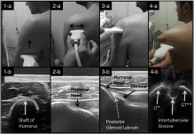
Figs. 1-4
Steps of performing a POCUS on shoulder; also defined in detail in Table 1 .
Fig. 5
Anterior (B) and posterior (C) glenohumeral dislocation and a postreduction view of the same patient with anterior dislocation (A). *PGL: posterior glenoid labrum.
Fig. 6
POCUS view of a normal humeral head and various types of fractures in shoulder. A normal humeral head, Hill-Sachs deformity, fracture of tuberculum majus, and a Bankart fracture are displayed in alphabetical order.
Abstract
Objective
Point-of-care ultrasonography (POCUS) is an easily available and noninvasive tool without radiation exposure that is also gaining a broad range of use in emergency departments. The aim of this study is to evaluate the value of POCUS in the diagnosis of shoulder dislocation by comparing with plain radiography.
Methods
This prospective observational study with a convenience sampling was conducted in emergency departments of 2 hospitals. Patients older than 15 years with possible shoulder dislocation during the physical examination composed the study population. All the study patients underwent POCUS evaluation to detect a shoulder dislocation or fracture before radiography, and the POCUS procedure was also achieved after the reduction attempt.
Results
A total of 103 patients were enrolled in the study. The mean age of study subjects was 33.9 ± 15 years, and 80.6% (n = 83) of them were male. The sensitivity and specificity of POCUS in identifying dislocation were 100% (95% confidence interval [CI], 96%-100%) and 100% (95% CI, 48%-100%), respectively. POCUS also confirmed reduction in 93 of 94 patients with a specificity of 100% (95% CI, 96%-100%). POCUS has a sensitivity of 100% (95% CI, 63%-100%) for excluding a shoulder fracture but a specificity of 84.2% (95% CI, 75%-91%).
Conclusion
Point-of-care ultrasonography is an effective tool to either rule in or rule out shoulder dislocation in the emergency setting. Furthermore, it is a robust sensitive tool for excluding fractures but with false-positive results.
To access this article, please choose from the options below
Purchase access to this article
Claim Access
If you are a current subscriber with Society Membership or an Account Number, claim your access now.
Subscribe to this title
Purchase a subscription to gain access to this and all other articles in this journal.
Institutional Access
Visit ScienceDirect to see if you have access via your institution.
☆Contributorship: collected data: Can Akyol, Faruk Gungor, Angelika Janitzky Akyol, Mustafa Kesapli, Ramazan Guven, Umut Cengiz, Halil İbrahim Toksul; served as scientific advisor: Eken C; statistical study: Eken C; writing the article: Eken C.
☆☆Funding: This research received no specific funding.
☆☆☆Competing interests: There are no competing interests
