


 Article Info
To view the full text, please login as a subscribed user or purchase a subscription. Click here to view the full text on ScienceDirect.
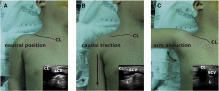
Fig. 1
The influence of different arm positions on the relative position of the clavicle the neutral position (A), caudal traction (B), and abduction (C). Abbreviations: CL, clavicle; L, lung.
Fig. 2
A, The long-axis in-plane view of the subclavian vein. B, Landmarks for the measurements. Abbreviations: CL, clavicle; FR, first rib; P, the point at which the lateral border of the IJV and the upper border of the SCV meet (Pirogoff confluence); S, the point at which the skin surface connects the vertical line tangentially drawn from the medial border of the clavicle; LL, the length of puncturable SCV; D, the diameter of the SCV at point P; α, the available angle of needle insertion.
Fig. 3
Ultrasound images of the SCV in 3 different arm positions the neutral position (A), caudal traction (B), and abduction (C). Abbreviations: LL, puncturable length of the SCV; D, diameter of the SCV; α, available angle for needle insertion. LL and α increase with caudal traction of the arm and decrease with abduction.
Fig. 4
Degree of increase (percentages) in the puncturable length of the SCV with caudal traction of the arm according to age. No significant difference between groups. Data are presented as means ± SD. The dots indicate each percent change in the length of the SCV.
Abstract
Background
The first step for successful ultrasound (US)–guided subclavian vein (SCV) catheterization using a supraclavicular approach is to obtain a good longitudinal image of SCV for in-plane needle placement. We evaluated the efficacy of caudal traction of ipsilateral arm on the exposure of the SCV.
Methods
We enrolled 20 infants, 20 children, and 20 adults undergoing general anesthesia. After tracheal intubation, US probe was applied as the supraclavicular approach, and the longitudinal US image of SCV was obtained in 3 different ipsilateral arm positions: neutral, caudal traction, and abduction. The length of puncturable SCV, the diameter of SCV, and the available angle for needle insertion in 3 different arm positions were analyzed.
Results
In all patients, the length of puncturable SCV and the available angle for needle insertion were significantly increased after caudal traction (35.6% ± 27.1% and 25.0% ± 19.3%, respectively) and decreased after the abduction (36.6% ± 22.9% and 29.5% ± 23.8%, respectively) compared to neutral position. The diameter of SCV was not changed after applying the caudal traction in infants and children. However, in adults, the caudal traction slightly increased the diameter of SCV (P = .012).
Conclusion
The caudal traction of ipsilateral arm toward to the knee improves the longitudinal US view of SCV for the supraclavicular approach, without reducing its size. Proper caudal traction of the arm might ensure the high success rate with safe needle insertion technique. Abduction should be avoided during US-guided supraclavicular SCV catheterization.
To access this article, please choose from the options below
Purchase access to this article
Claim Access
If you are a current subscriber with Society Membership or an Account Number, claim your access now.
Subscribe to this title
Purchase a subscription to gain access to this and all other articles in this journal.
Institutional Access
Visit ScienceDirect to see if you have access via your institution.
☆Conflict of interest: The authors have no conflict of interest.
☆☆Funding: Financial support for the study was provided solely from departmental source.
