Resuscitating the tracheostomy patient in the ED
Affiliations
- Department of Emergency Medicine, San Antonio Military Medical Center, Houston, TX 78234
Correspondence
- Corresponding author at: 506 Dakota St APT 1, San Antonio, TX 78203. Tel.: +1 719 339 5510.

Affiliations
- Department of Emergency Medicine, San Antonio Military Medical Center, Houston, TX 78234
Correspondence
- Corresponding author at: 506 Dakota St APT 1, San Antonio, TX 78203. Tel.: +1 719 339 5510.
Affiliations
- Department of Emergency Medicine, The University of Texas Southwestern Medical Center, Dallas, TX 75390
 Article Info
To view the full text, please login as a subscribed user or purchase a subscription. Click here to view the full text on ScienceDirect.

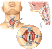
Fig. 1
Normal location and anatomy of tracheostomy site. Image from http://biology-forums.com/index.php?action=gallery;sa=view;id=10069.
Fig. 2
Bjork flap with anatomy of tracheostomy site. Image from http://pocketdentistry.com/12-surgical-management-of-the-airway/.
Fig. 3
Tracheostomy components. Image from https://patienteducation.osumc.edu/documents/fenestr.pdf.
Fig. 4
Airway management strategy.
Fig. 5
Algorithm for management of tube obstruction and dislodgement.
Fig. 6
Algorithm for managing the bleeding tracheostomy site.
Fig. 7
Tracheostomy balloon inflation for bleeding. Hyperinflate balloon to 35 to 50 mL. Image from http://www.downstatesurgery.org/files/cases/tif.pdf.
Fig. 8
External and internal manual compression of stoma site with oral intubation. Image from http://www.downstatesurgery.org/files/cases/tif.pdf.
Abstract
Background
Emergency physicians must be masters of the airway. The patient with tracheostomy can present with complications, and because of anatomy, airway and resuscitation measures can present several unique challenges. Understanding tracheostomy basics, features, and complications will assist in the emergency medicine management of these patients.
Objective of review
The aim of this review is to provide an overview of the basics and features of the tracheostomy, along with an approach to managing tracheostomy complications.
Discussion
This review provides background on the reasons for tracheostomy placement, basics of tracheostomy, and tracheostomy tube features. Emergency physicians will be faced with complications from these airway devices, including tracheostomy obstruction, decannulation or tube dislodgement, stenosis, tracheoinnominate fistula, and tracheoesophageal fistula. Critical patients should be evaluated in the resuscitation bay, and consultation with ENT should be completed while the patient is in the department. This review provides several algorithms for management of complications. Understanding these complications and an approach to airway management during cardiac arrest resuscitation is essential to optimizing patient care.
Conclusion
Tracheostomy patients can present unique challenges for emergency physicians. Knowledge of the basics and features of tracheostomy tubes can assist physicians in managing life-threatening complications including tube obstruction, decannulation, bleeding, stenosis, and fistula.
To access this article, please choose from the options below
Purchase access to this article
Claim Access
If you are a current subscriber with Society Membership or an Account Number, claim your access now.
Subscribe to this title
Purchase a subscription to gain access to this and all other articles in this journal.
Institutional Access
Visit ScienceDirect to see if you have access via your institution.
Conflicts of interest: None.
This clinical review has not been published, it is not under consideration for publication elsewhere, its publication is approved by all authors and tacitly or explicitly by the responsible authorities where the work was carried out, and that, if accepted, it will not be published elsewhere in the same form, in English or in any other language, including electronically without the written consent of the copyright holder.
Related Articles
Searching for related articles..


