Article, Orthopedics

The effectiveness of a newly developed reduction method of anterior shoulder dislocations; Sool’s method

a b s t r a c t

Objective: Nearly a dozen Reduction methods for the treatment of anterior Shoulder dislocation have been report- ed, but the majority are painful and require patients to be in the supine or prone position.

Methods: This retrospective cohort study was conducted in a university-affiliated emergency department (ED). Sool’s method and traditional shoulder reduction methods (TSRMs) were performed for the patient with Anterior shoulder dislocation. Fifty-nine eligible patients were recruited; 35 were treated with TSRMs, wherease 24 were treated with Sool’s method.

Results: The rate of Successful reduction was 80% (26/35) in the TSRM group and 75% (18/24) in the Sool’s method group (P = .75). The length of stay in the ED was 72.3 minutes in the Sool’s method group and 98.4 minutes in the TSRM group (P = .037). No significant difference was observed between the neurovascular deficit before and after reduction in either group. The procedural time of successfully reduced cases in patients treated by Sool’s method was shorter than that of the failed cases (P = .015).

Conclusions: Sool’s method was as successful as other methods at reducing shoulder dislocation and has demon- strated encouraging results, including significant reduction in length of stay in the ED and unnecessary use of se- dation. Sool’s method is technically easy and requires only a place to sit and a single operator.

(C) 2016

Introduction

Shoulder dislocations are classified according to their etiology, the direction of instability, or combinations thereof [1]. Anterior shoulder dislocation, which accounts for nearly 90%-95% of all shoulder disloca- tions, is a common complaint in emergency departments (EDs) [2].

Numerous methods for anterior shoulder reduction have been re- ported [3]. These techniques can be divided into 4 categories using 4 main headings of traction: countertraction, leverage, scapular manipu- lation, and various combinations of these maneuvers. Some traditional shoulder reduction methods (TSRMs) are technically difficult to per- form and have other disadvantages [4]. Many versions of TSRM require that the patient lie on a bed to reduce the dislocated Shoulder joint.

? Funding source: The authors have no financial relationships relevant to this article to disclose.

?? Financial disclosure: The authors have nothing to disclose.

??? Conflict of interest: The authors have no conflicts of interest to disclose.

? Copyright transfer: The authors consented to the copyright transfer agreement.

?? Contributors’ statement: Moon Seok Park, Jin Hee Lee: Dr Park and Dr Lee equally

conceptualized and designed the study, drafted the initial manuscript, and approved the

final manuscript as submitted.

* Corresponding author at: Department of Emergency Medicine, Seoul National Uni- versity Bundang Hospital, 300 Gumi-dong, Bundang-gu, Seongnam-si, Gyeonggi-do, 463-707, Korea. Tel.: +82 10 3424 6718; fax: +82 31 787 4081.

E-mail address: [email protected] (H. Kwon).

Lying on a bed is often uncomfortable, and some patients cannot per- form this because of the pain involved. In the study presented herein, we describe a new and easy method for shoulder reduction that reduces pain and provides and alternative position; this method is known as Sool’s method.

The goal of this study was to introduce Sool’s method and assess the efficacy of this newly described technique.

Methods

This study was performed at an urban tertiary care hospital with 1100 beds. The hospital reports about 80,000 ED visits annually. A retro- spective medical record review of patients presenting to the ED with an- terior shoulder dislocation between January 2012 and August 2014 was conducted to compare Sool’s method of shoulder reduction with other TSRMs. Patients in the TSRM group underwent versions of Kocher, Spaso, Hippocratic traction with countertraction, and Stimson methods for shoulder reduction.

Patients who were documented both clinically and radiographically with an acute anterior shoulder dislocation at the time of initial presen- tation at the ED were included in this study. Neurovascular status was also documented. Patients with hemodynamic instability, multiple traumas, general hyperlaxity, dislocations associated with severe

http://dx.doi.org/10.1016/j.ajem.2016.04.012

0735-6757/(C) 2016

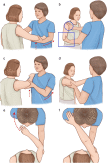
Figure. Sequence of Sool’s method for reduction of anterior shoulder dislocation.

glenoid fractures, a history of previous shoulder surgery, and disloca- tions associated with humeral fractures were excluded. Patients who presented more than 24 hours after the injury were also excluded.

This study was approved by the Institutional Review Board at Seoul National University Bundang Hospital. Informed consent was waived because of the retrospective nature of the study.

Table 1

Baseline characteristic between TSRM and the Sool’s method groups

Table 3

Comparison of successful and failed cases of the Sool’s method

TSRM n = 35

Sool’s method

P value

Successful reduction

Failed reduction

P value

n = 24

n = 18

n = 6

Age (median, IQR)

35 (22~ 52)

39.5 (21.5 ~ 65)

.53

Age (median, IQR)

28.5 (20~ 60)

66 (46~ 82)

.016

Male, n (%)

27 (77)

13 (54)

.09

Male, n (%)

11 (61)

2 (33)

.36

Traumatic, n (%)

14 (40)

14 (58)

1.00

Procedure duration

14 (78)

1 (17)

.015

Neurovascular status before reduction (b 2 min), n (%)

Intact, n (%)

31 (89)

21 (87)

1.00

Deficit, n (%)

4 (11)

3 (13)

1.00

Neurovascular status after reduction

Intact, n (%)

34 (97)

24 (100)

1.00

Deficit, n (%)

1 (3)

0 (0)

1.00

Initial method outcome

Successful reduction, n (%)

26 (80)

18 (75)

.75

Recurrent dislocation, n (%)

11 (31)

6 (25)

.77

Successful initial reduction, n (%)

8 (73)

4 (67)

1.00

LOS (min), mean (95% CI)

98.4 (80.5 ~ 116.3)

72.3 (58.3 ~ 86.2)

.037

IQR, interquartile range; CI, confidence interval.

Development of Sool’s method

patient comfort and relaxation are key factors for successful shoul- der reduction [5]. The authors of this study proposed that dislocation of the shoulder joint may cause spasm of muscles, including the pectoralis and deltoids, which could result in difficult reduction of the dislocated joint.

The authors focused on massaging techniques that increase muscle compliance, resulting in an increased range of joint motion, decreased passive spasm, and decreased active spasm (biomechanical mecha- nisms) [6]. The authors compared patients treated with TSRMs with those treated with Sool’s method. The final version of the maneuver was reported in December 2012. This method has been used in the ED since 2013. “Sool” was named after author Dr Hyuksool Kwon, who was the first to propose this method.

Sequence of Sool’s method (Figure; see Video, Supplemental Digital Content 1, which demonstrates the successful cases of Sool’s method)

The patient is placed in a sitting position. The operator asks the patient to straighten their back and not to rotate their trunk for the duration of the procedure.
  • The operator stands in front of the patient.
  • The operator asks the patient to raise the affected arm and hand with forward flexion until the angle of the arm and trunk reaches 90?. The patient’s hand is placed on the operator’s shoulder of the same side (eg, if the patient’s dislocated shoulder is on their right side, the patient’s right hand is placed on the operator’s right shoul- der). If pain occurs, the operator stops the maneuver and waits until the pain subsides before lifting the arm again (Figure A).
  • The operator grasps the anterior part of the deltoid and pectoralis muscles from the patient’s affected side with their hand on the same side (eg, if the patient’s dislocated shoulder is on their right side, the operator touches the anterior part of the deltoid and pectoralis with the operator’s right hand) (Figure B, E, and F).
  • The operator grasps the elbow of the affected side with the oppo- site hand.
  • The operator slowly pulls the elbow and massages the anterior part of the deltoid and pectoralis muscles. The operator asks the patient to breathe slowly and deeply to relax the muscles and em- phasizes straightening their back (Figure C and D).
  • When the operator feels a clunk, the reduction is complete. After the reduction, the location of the humeral head is confirmed both clinically and radiographically.
  • Patients are not allowed to move their shoulder into a position of adduction and internal rotation for 3 weeks; this is done with a shoulder immobilizer.
  • Sool’s method was first performed February 20, 2013. Patients diag- nosed with anterior shoulder dislocation were treated by emergency physicians trained in this method. When a patient’s shoulder failed to be reduced by Sool’s method during the initial attempt or if a patient re- quested sedation for reduction, they were treated using TSRMs.

    With respect to patients in the TSRM group, physicians were allowed to use any method for closed reduction according to their preference and clinical judgment because no single shoulder reduction method has been proven to be superior to other methods. Morphine (>= 2 mg) was allowed in patients in the TSRM group before reduction. In addition, intravenous etomidate or ketamine could be used before shoulder re- duction if deemed necessary by the physician. Patients in the Sool’s method group were not given analgesics or sedatives initially.

    Outcome measures

    The primary outcome measure for this study was successful reduc- tion rate of the first attempt at reduction. Secondary outcome measures included length of patient stay (LOS) in the ED, complication rate before and after reduction, sedation rate of the failed first attempt of reduction, and length of the procedure.

    Data analysis

    The Stata version 13.1 statistical software (Stata Corp LP) was used for data analysis. Data are expressed as either means +- SD or medians with interquartile range. Continuous variables were compared using Student t test for independent samples with normal distributions. Non- parametric analysis was performed for continuous data that did not fol- low a normal distribution. Fisher exact test was used for categorical data distribution. All P values were 2-tailed, and a P value b .05 was consid- ered to indicate statistical significance.

    Table 2

    Table 4

    Comparison of failed cases of initial Reduction attempt between TSRM and Sool’s method

    Comparison of successful cases

    between the Sool’s me

    thod and TSRM

    TSRM

    Sool’s method

    P value

    Sool’s method

    TSRM

    P value

    n = 9

    n = 6

    n = 18

    n = 26

    Age (median, IQR)

    25 (19~ 58)

    66 (46~ 82)

    .14

    Age (median, IQR)

    24 (20-46)

    35 (26-45)

    .333

    Male, n (%)

    7 (78)

    2 (33)

    .14

    Male, n (%)

    13 (72)

    20 (77)

    .738

    Sedation for further reduction

    3 (33)

    4 (66)

    .32

    LOS (min), mean (95% CI)

    55.2 (51.5-59.0)

    84.0 (68.9-99.2)

    b.001

    attempt, n (%)

    Table 5

    Summary of relocation method of anterior shoulder dislocation

    Procedure

    Success rate

    Advantage

    Disadvantage

    Leverage

    Kocher’s method

    Bend the affected arm at 90? at the

    100%

    Highly successful

    This method can cause fractures

    elbow, adduct against the body

    of the Proximal humerus or

    Milch technique

    Abduct and externally rotate the

    89.4%

    This reduction is tolerated

    neurovascular compromise.

    No significant disadvantage

    External rotation

    patient’s arm into an overhead position

    Adduct, bend affected elbow at 90?,

    80%

    well by patients. Procedural sedation is not necessary.

    A single operator and minimal force are required.

    This reduction is tolerated

    Success rate may be lower than

    Traction

    gently externally rotate the forearm

    well by patients.

    A single operator and no force or traction are required. Procedural sedation is not necessary.

    The reduction can be done quickly and easily.

    other methods.

    Hippocratic method

    Stimson’s method

    Place the heel of the practitioner in the axilla (not pressed hard), adduct, and pull the arm

    The patient prone on a table with

    Not reported

    96%

    Highly successful

    No assistance is required.

    This method can cause fractures of the proximal humerus or neurovascular compromise.

    The patient must be monitored

    the affected arm hanging down in forward flexion with 10-lb weight

    The shoulder is reduced with minimal force (gravity and

    weights).

    at all times.

    Equipment and sufficient premedication are necessary.

    Traction-countertraction

    Traction applied to the affected

    92%

    Procedure is useful in patients

    The time required for reduction is relatively long.

    Sedation and more than 1 operator

    Spaso technique

    arm while the shoulder is in abduction, countertraction to the chest

    Lift and externally rotate the

    87.5%

    with severe Muscle spasm or pain and in those who cannot relax.

    A single operator is required.

    are required.

    This reduction requires prolonged force and endurance.

    Equipment is needed.

    No significant disadvantage

    Chair method

    shoulder, then push the humeral head back

    Sit in a stable chair sideways, using

    100%

    Minimal force is required.

    A single operator and no

    The need for an appropriate chair

    the backrest of the chair as a fulcrum in the axilla

    force or traction are required. Procedural sedation is not necessary.

    The reduction can be done

    quickly and easily.

    which may not always be available in certain circumstances.

    Scapular manipulation

    Sitting

    Place the affected arm in 90? of

    90.2%

    This reduction is tolerated

    The borders of the scapula are

    Supine

    forward flexion at the shoulder, and slight traction.

    Rotate the inferior tip of the scapula medially and the superior

    aspect laterally with slight dorsal

    100%

    well by patients.

    Reduction can be performed without premedication.

    Minimal force is required.

    difficult to locate in obese patients.

    Assistance is needed for traction

    if the patient is prone and weights are unavailable or if the patient is seated.

    displacement.

    Results

    During the data collection period from January 2012 to August 2014, 59 eligible patients who presented to the ED were recruited. Physicians were allowed to decide whether to perform TSRMs or Sool’s method. Thirty-five patients were treated by TSRMs, whereas 24 patients were treated by Sool’s method.

    In the Sool’s method group, reduction failed in 6 patients (25%), who proceeded to undergo an additional reduction using TSRMs. No patient failed in the final reduction. The 2 treatment groups had similar baseline characteristics (Table 1). The primary outcome measure of the rate of successful reduction of the initial attempt was 80% (26/35) in the TSRM group and 75% (18/24) in the Sool’s method group (P = .75). With regard to secondary outcomes, the LOS in the ED was

    72.3 minutes in the Sool’s method group and 98.4 minutes in the TSRM group (P = .037) (Table 1). No adverse event in either reduction or sedation was reported. There was no significant difference in neurovascular deficit before and after reduction in either group. In those reduction cases that were successful at the initial attempt, the

    LOS was significantly less for patients in the Sool’s method group than those in the TSRM group (Table 2).

    When comparing successful cases between Sool’s method (Table 3), the procedural time of successfully reduced cases was shorter (less than 2 minutes) than failed cases (P = .015). Patients were significantly older in failed cases (P = .016). Sixty-six percent (4/6) of failed cases from the Sool’s method group required sedation before further reduc- tion with other TSRMs, whereas 33% (3/9) of failed cases with initial TSRMs required sedation before further reduction attempts (Table 4).

    Discussion

    Sool’s method, a newly developed reduction method for the treat- ment of anterior shoulder dislocations, reduced LOS in the ED success- fully without significantly decreasing the rate of reduction. We hypothesized that overcoming muscle spasms and reducing pain for re- laxation would be paramount for a successful reduction. To achieve ad- equate patient relaxation, we chose to massage the shoulder muscle group and placed patients in a seated position.

    Sool’s method is easy to perform, effective, free of complications, and easily applicable (see Video, Supplemental Digital Content 1, which demonstrates the successful cases of Sool’s method). It is also rapid, as 78% of dislocated shoulders were reduced within 2 minutes (Table 3). The success rate of this new method was 75% (Table 1), similar to the success rates of various reduction methods for treating anterior shoul- der Joint dislocations, which range from 70% to 100% [7-11]. Sool’s method does not require sedation or an assistant, which greatly affects the LOS in the ED (Tables 1 and 2). The seated position eliminates pain and discomfort associated with lying on a bed in the prone or supine po- sition. Furthermore, massaging the deltoid and pectoralis muscles both enhances relaxation of spasmodic muscles and facilitates reduction.

    Shoulder function can be impaired by muscle, bone, and/or nerve in- juries after reduction [12]. Approximately 10% of patients suffer injuries to the Axillary nerve following anterior dislocation [13]. No nerve, mus- cle, or bone injuries were observed in this study. The lack of complica- tions confirms the safety of this method (Table 1).

    We showed that 66% (4/6) of failed cases of Sool’s method required

    sedation before reduction by other TSRMs (Table 4). Moreover, all pa- tients underwent several rounds of reduction with other TSRMs after failing Sool’s method, suggesting that reduction by TSRM without seda- tion could be difficult even if it was treated by TSRM initially and not only Sool’s method.

    Various methods for anterior shoulder dislocation reductions have been reported (Table 5), each of which has advantages and disadvan- tages. The Hippocratic method and Kocher maneuver are not recom- mended because of the high incidence of axillary nerve injury, capsular damage, and humeral shaft and neck fractures [14,15]. Scapu- lar manipulation techniques are difficult to apply to overweight persons and require patients to lie in the prone position [16]. The Stimson tech- nique requires a long time and is not well tolerable to patients [17]. Spaso’s technique involves traction and external rotation; traction in- creases muscle spasms, and excessive external rotation may cause a hu- meral fracture [18,19]. The Milch technique is a complex maneuver [20]. Chair method needs an appropriate chair which may not always be available in certain circumstances [21]. Sool’s method is performed in a seated position with assisted forward elevation of the affected arm, and relaxes muscles through massaging, which may reduce muscle spasms and muscle, nerve, and bone injuries during reduction.

    Sool’s method has several drawbacks inferred from the failed cases. First, this cannot be applied to a patient who cannot sit in a straight line. Bending or rotating of the patient’s torso interrupts the appropriate position and disturbs successful reduction. Reduction with Sool’s meth- od was especially difficult in elderly patients who could not sit in a straight line for a longer procedural duration (more than 2 minutes) (Table 3). Second, this method cannot be attempted if a patient is un- conscious or unable to raise the affected arm and hand because of severe pain. Third, for a successful and Rapid reduction, the physician needs enough strength for grasping, traction, and massaging of the affected arm.

    This study has several limitations. First, this was retrospective study with a nonblinded medical record review. Authors may be biased by the use of their own technique and not being blinded to data collection. Sec- ond, the small sample size likely affected the statistical results, especial- ly the difference between failed reduction rate according to sex or age.

    Third, Sool’s method was conducted by the authors at a single center. Therefore, the feasibility and repeatability of this new method could not be accessed. Fourth, no patient received follow-up; therefore, de- layed complications or recurring dislocations could not be considered in this study. A planned, prospective study would further confirm these results, particularly addressing any limitations within this study.

    Conclusions

    Sool’s method is technically easy and requires only a place to sit and a single operator, which reduces the use of valuable ED resources. Sool’s method has demonstrated encouraging results, including significant re- duction in LOS at the ED and unnecessary use of sedation.

    Supplementary data to this article can be found online at http://dx. doi.org/10.1016/j.ajem.2016.04.012.

    References

    1. Gerber C, Nyffeler RW. Classification of Glenohumeral joint instability. Clin Orthop Relat Res 2002:65-76.
    2. Zacchilli MA, Owens BD. Epidemiology of shoulder dislocations presenting to emer- gency departments in the United States. J Bone Joint Surg Am 2010;92:542-9.
    3. Gleeson AP. Anterior glenohumeral dislocations: what to do and how to do it. J Accid

      Emerg Med 1998;15:7-12.

      Riebel GD, McCabe JB. Anterior shoulder dislocation: a review of reduction tech- niques. Am J Emerg Med 1991;9:180-8.

    4. Canales Cortes V, Garcia-Dihinx Checa L, Rodriguez VJ. Reduction of acute anterior dislocations of the shoulder without anaesthesia in the position of maximum mus- cular relaxation. Int Orthop 1989;13:259-62.
    5. Weerapong P, Hume PA, Kolt GS. The mechanisms of massage and effects on perfor- mance, muscle recovery and injury prevention. Sports Med 2005;35:235-56.
    6. Singh S, Yong CK, Mariapan S. Closed reduction techniques in acute anterior shoul- der dislocation: modified Milch technique compared with traction-countertraction technique. J Shoulder Elbow Surg 2012;21:1706-11.
    7. Caudevilla Polo S. Estebanez de Miguel E, Lucha Lopez O, Tricas Moreno JM, Perez Guillen S. Humerus axial traction with acromial fixation reduction maneuver for an- terior shoulder dislocation. J Emerg Med 2011;41:282-4.
    8. Kothari RU, Dronen SC. Prospective evaluation of the scapular manipulation tech- nique in reducing anterior shoulder dislocations. Ann Emerg Med 1992;21:1349-52.
    9. Beattie TF, Steedman DJ, McGowan A, Robertson CE. A comparison of the Milch and Kocher techniques for acute anterior dislocation of the shoulder. Injury 1986;17: 349-52.
    10. Thakur AJ, Narayan R. Painless reduction of shoulder dislocation by Kocher’s meth- od. J Bone Joint Surg Br 1990;72:524.
    11. Johnson JR, Bayley JI. early complications of acute anterior dislocation of the shoul- der in the middle-aged and elderly patient. Injury 1982;13:431-4.
    12. Visser CP, Coene LN, Brand R, Tavy DL. The incidence of nerve injury in anterior dis- location of the shoulder and its influence on functional recovery. A prospective clin- ical and EMG study. J Bone Joint Surg Br 1999;81:679-85.
    13. Marx J, Hockberger R, Walls R. Rosen’s emergency medicine-concepts and clinical practice: Elsevier health sciences; 2013.
    14. Christofi T, Kallis A, Raptis D, Rowland M, Ryan J. Management of shoulder disloca- tions. Trauma 2007;9:39-46.
    15. Sahin N, Ozturk A, Ozkan Y, Atici T, Ozkaya G. A comparison of the scapular manip- ulation and Kocher’s technique for acute anterior dislocation of the shoulder. Eklem Hastalik Cerrahisi 2011;22:28-32.
    16. O’Connor DR, Schwarze D, Fragomen AT, Perdomo M. Painless reduction of acute an- terior shoulder dislocations without anesthesia. Orthopedics 2006;29:528-32.
    17. Uglow MG. Kocher’s painless reduction of anterior dislocation of the shoulder: a pro- spective randomised trial. Injury 1998;29:135-7.
    18. Bucholz RW. Rockwood and Green’s fractures in adults: Lippincott Williams & Wilkins Philadelphia; 2006.
    19. Lacey II T, Crawford HB. Reduction of anterior dislocations of the shoulder by means of the Milch abduction technique. J Bone Joint Surg Am 1952;34-a:108-9.
    20. Mahirogullari M, Akyildiz F, Koksal I, Cakmak S, Kurklu M, Kuskucu M. Chair method: a simple and effective method for reduction of anterior shoulder dislocation. Acta Orthop Traumatol Turc 2012;46:102-6.