


Abstract
Objectives
To establish a standardized approach for the rapid and accurate identification of
non-traumatic, ophthalmologic pathology in patients with eye complaints in the emergency
department.
Methods
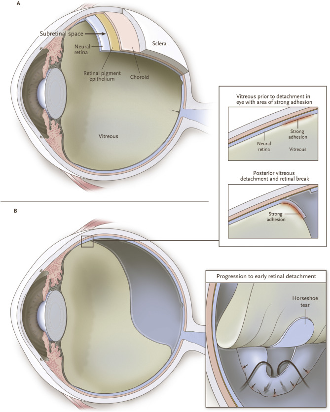
In this detailed protocol we offer an easy, reproducible method for the use of ocular
point-of-care ultrasound (POCUS) in helping practitioners identify and distinguish
between common eye pathology encountered in the emergency setting: retinal detachment,
vitreous detachment, vitreous hemorrhage, optic nerve pathology, and syneresis.
Conclusions
This protocol can help identify patients that may need urgent ophthalmology consultation
those that can follow-up on an outpatient, and those that may need additional emergent
testing.
Keywords
To read this article in full you will need to make a payment
One-time access price info
- For academic or personal research use, select 'Academic and Personal'
- For corporate R&D; use, select 'Corporate R&D; Professionals'
Subscribe:
Subscribe to The American Journal of Emergency MedicineAlready a print subscriber? Claim online access
Already an online subscriber? Sign in
Register: Create an account
Institutional Access: Sign in to ScienceDirect
References
- How effective is undergraduate and postgraduate teaching in ophthalmology?.Eye. 1997; 11 (September): 744-750
- Feasibility of nonmydriatic ocular fundus photography in the emergency department: phase I of the FOTO-ED study.Acad Emerg Med. 2011; 18 (Sep): 928-933
- Bedside ocular ultrasound for the detection of retinal detachment in the emergency department.Acad Emerg Med. 2010; 17 (Sep): 913-917
- A study of bedside ocular ultrasonography in the emergency department.Acad Emerg Med. 2002 Aug; 9: 791-799
- Elevated intracranial pressure detected by bedside emergency ultrasonography of the optic nerve sheath.Acad Emerg Med. 2003 Apr; 10: 376-381
- Point-of-care ocular ultrasound to detect optic disc swelling.Acad Emerg Med. 2013; 20 (Sep): 920-925
- Syneresis and swelling of gelatin.J Gen Physiol. 1928; 12 (Nov 20,): 289-312
- Adult vitreous structure and postnatal changes.Eye (Lond). 2008; 22 (Oct): 1214-1222
- Posterior vitreous detachment: evolution and complications of its early stages.Am J Ophthalmol. 2010 Mar; 149: 382.e1
. D'Amico DJ. Clinical practice. Primary retinal detachment. N Engl J Med. 2008 Nov 27;359(22):2346–54.
- Retinal detachment in Olmsted County, Minnesota, 1976 through 1995.Ophthalmology. 1999; 106 (Jan): 154-159
- Causes and treatment of vitreous hemorrhage.Compr Ophthalmol Update. 2006; 7 (May-Jun): 97-111
- Vitreoretinal disorders.Ultrasound Clinics. 2008; 3: 217-228
- Optic nerve ultrasound for the detection of raised intracranial pressure.Neurocrit Care. 2011; 15 (December 01): 506-515
- Ultrasonography of the optic nerve sheath may be useful for detecting raised intracranial pressure after severe brain injury.Intensive Care Med. 2007; 33 (October 01): 1704-1711
- Optic nerve sheath diameter and lumbar puncture opening pressure in nontrauma patients suspected of elevated intracranial pressure.Am J Emerg Med. 2014; 32 (December 01): 1513-1515
- Morphometry of the retrobulbar human optic nerve: comparison between conventional sonography and ultrafast magnetic resonance sequences.Invest Ophthalmol Vis Sci. 2007 May 01; 48: 1913-1917
- Accuracy of optic nerve sheath diameter measurement by emergency physicians using bedside ultrasound.J Emerg Med. 2015; 48 (April 01): 450-457
- Reproducibility and accuracy of optic nerve sheath diameter assessment using ultrasound compared to magnetic resonance imaging.BMC Neurol. 2013; 13 (December 01): 187
Article Info
Publication History
Published online: May 11, 2019
Accepted:
April 28,
2019
Received in revised form:
April 26,
2019
Received:
December 19,
2018
Identification
Copyright
© 2019 Elsevier Inc. All rights reserved.
