Article, Emergency Medicine

The pivotal role of a pre-admission electrocardiogram for the diagnosis of Takotsubo syndrome in the ED

Correspondence

Contents lists available at ScienceDirect

American Journal of Emergency Medicine

journal homepage: locate/ ajem

The pivotal role of a pre-admission electrocardiogram for the diagnosis of Takotsubo syndrome in the ED?

To the Editor,

Prompt differentiation of Takotsubo syndrome (TTS) from acute coronary syndromes is crucial in the management of patients presenting with chest pain/dyspnea/palpitations/weakness/dizziness/ presyncope/syncope in the emergency department (ED), since it is imperative that the latter receive expedited therapy with thrombolysis or Percutaneous coronary interventions, while the former are cared for along the lines of symptomatic nonspecific management, until we know more about the pathogenesis of TTS [1]. The crux of the problem at hand is whether we possess a noninvasive diagnostic tool for the differenti- ation of the above 2 conditions, or we need invasive coronary arteriography to tell them apart. Indeed even with coronary arteriogra- phy half of the patients with eventually documented “normal or unobstructed coronary arteries” in a recent series [1] remained undiagnosed since a contrast ventriculogram was not done at the time of cardiac catheterization. This underscores the importance of doing a contrast ventriculogram, or performing a transthoracic echocardiogram

either immediately after cardiac catheterization, or (even better) at the ED, before this invasive procedure. The quest for a noninvasive Diagnostic modality, or index to differentiate TTS from ACS goes on.

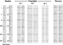
In a recent communication, published in the Journal [2], this author outlined a diagnostic scenario for physicians working in EDs, based on the merits of the electrocardiogram (ECG), which could be implemented immediately upon encountering a patient who could have suffered a case of TTS. This usually is a postmenopausal woman in her 60s with or without history of hypertension, with her first complains of symptoms suggestive of an ACS, or a history of previous such incidences with an eventual negative work up for an ACS. It has been reported recently that patients with TTS characteristically show in their ECGs attenuation of the QRS voltage (AQRSV), particularly in Lead aVR, all limb leads, and leads V4 to V6 [3-6]. The admission ECG acquires the features of low voltage ECG (QRS complexes <=5 mm in limb leads, and <=10 mm in the precordial leads) or show marked AQRSV in comparison with ECGs obtained prior to the index admission. Thus it is imperative that managing physicians obtain such previous ECG(s) for comparison, merely glancing at them, set side by side [2]. Since a picture is worth a thousand words, here is an example from the literature (Fig.) [7], for the readers’ scrutiny, of 5 ECGs of a 45-year-old woman with TTS, obtained 11 months before presentation, on admission, days 3 and 5 of hospitalization, and 5 weeks after presentation, during

Fig. Analysis of the ECG as in the text. (Reproduced from Fig. 1 of Ref. 7, with the permission of Elsevier).

? Conflict of interest: None declared.

0735-6757/(C) 2016

recovery. The AQRSV of the admission ECG in comparison with her previous ECG is apparent. Although further AQRSV occurred in the ECGs from days 3 and 5, the admission ECG showed already AQRSV in comparison with the previous ECG of the patient, although she presented only a few hours after the “sudden onset of her acute distress” [7]. Thus, the admission ECG can be used in the ED for the diagnosis of TTS. This will eventually be either corroborated or refuted by the experiences of ED physicians.

John E. Madias, MD

Icahn School of Medicine at Mount Sinai

New York, NY, USA Division of Cardiology

Elmhurst Hospital Center, Elmhurst

NY 11373, USA

E-mail address: [email protected] http://dx.doi.org/10.1016/j.ajem.2013.12.002

References

  1. Sharma V, Srinivasan M, Sheehan DM, Ionescu A. stress cardiomyopathy: case series and the review of literature. J Emerg Med 2013;45:e95-8.
  2. Madias JE. Voltage attenuation of the electrocardiogram in Takotsubo syndrome: what ED physicians should consider. Am J Emerg Med 2014;32:102.
  3. Madias JE. Electrocardiogram in myocardial edema due to Takotsubo syndrome. J Electrocardiol 2012;45:795-6.
  4. Madias JE. Electrocardiogram lead-specific QRS attenuation in an atypical midventricular case of Takotsubo syndrome. J Electrocardiol. 2013 Sep 9. pii: S0022-0736(13)00414-7. http://eresources.library.mssm.edu:2099/10.1016/j. jelectrocard.2013.08.008. [Epub ahead of print].
  5. Madias JE. Electrocardiogram repolarization changes in patients with Takotsubo and acute coronary syndromes. American Journal of Cardiology (in press).
  6. Madias JE. Transient attenuation of the amplitude of the QRS complexes in the diagnosis of Takotsubo syndrome. Eur Heart J: acute cardiovascular care (in press).
  7. Donker DW, Pragt E, Weerwind PW, Holtkamp JW, Vainer J, Mochtar B, et al. Rescue Extracorporeal life support as a bridge to reflection in fulminant stress-induced cardiomyopathy. Int J Cardiol 2012;154:e54-6.

    A Letter to the editor: carbon monoxide poisoning deaths in the United States, 1999 to 2012

    To the Editor,

    We read the article with great interest by Sircar et al, which characterized unintentional, non-fire-related (UNFR) Carbon monoxide poisoning deaths in the United States [1]. We commend the authors’ efforts in their attempt to identify at-risk populations and risk stratify patients based on demographic factors. This information would be vital to prevention.

    Several demographic factors were noted to be significant in the UNFR CO deaths: male sex, marital status, age, race, and location. Figure 1 visually identifies the states with the highest occurrence of UNFR CO deaths. We point out that it would have been useful to include a similar map reflecting temporal trends in CO detector laws in each state as a possible explanation for the geographic discrepancy. In addition, the authors of the study do not include carboxyhemoglobin (COHb) concentrations in any of the fatalities. Carbon monoxide is detectable postmortem [2], and COHb concentrations would have been useful to compare among the varying populations. Because victims older than 85 years were at the highest risk for fatality, if preexisting medical conditions commonly seen in the elderly predispose them to CO toxicity, the degree of CO poisoning based on COHb concentrations could have

    accounted for the increased mortality in the elderly population.

    In conclusion, the article by Sircar et al examines an important question regarding UNFR CO deaths. The investigation of the CO detector laws could have greatly supported the conclusion of the authors. Without specifically examining this issue, it is not possible to draw a significant conclusion regarding CO deaths in relation to CO detector use. Furthermore, knowledge of COHb values could have assisted in explaining differences in CO-related mortality.

    Daria Falkowitz, DO

    North Shore University Hospital, North Hempstead, NY

    Corresponding author

    E-mail address: [email protected]

    Stephen Alerhand, MD

    Icahn School of Medicine at Mount Sinai, New York, NY

    Mark Su, MD, MPH The Ronald O Perelman Department of Emergency Medicine New York University School of Medicine, New York, NY

    http://dx.doi.org/10.1016/j.ajem.2016.01.011

    References

    Sircar K, Clower J, Shin MK, Bailey C, King M, Yip F. Carbon monoxide poisoning deaths in the United States, 1999 to 2012. Am J Emerg Med 2015;33(9):1140-5.

  8. Yonemitsu K, Sasao A, Oshima T, Mimasaka S, Ohtsu Y, Nishitani Y. Quantitative

    evaluation of volatile hydrocarbons in post-mortem blood in forensic autopsy cases of fire-related deaths. Forensic Sci Int 2012;217(1-3):71-5.

    Hemodynamic challenges in traumatic subarachnoid hemorrhage complicated by cerebral vasospasm

    To the Editor,

    We have read with great interest the recent review by Varvarousi et al [1] on the role of levosimendan in the clinical setting of Aneurysmal subarachnoid hemorrhage (SAH). Previous work by us [2] and others [3,4] proposed depressed cardiac output (CO), mainly attributed to catecholamine-mediated myocardial dysfunction, as a critical determinant of delayed cerebral ischemia (DCI) and Poor neurologic outcome. In this respect, rapid hemodynamic augmenta- tion, with either levosimendan [5] or other agents [6], has the potential to reverse cerebral ischemia and prevent further brain damage. However, analogous data are sparse in the setting of Traumatic SAH. We present herein the clinical course of 2 patients with traumatic SAH and cardiovascular derangement and emphasize the potential influence of early CO optimization and levosimendan administration in preventing delayed neurologic complications.

    A 70-year-old man with traumatic SAH (Figure A) was intubated with Glasgow Coma Scale (GCS) of 8/15. At the time of admission in the intensive care unit, he was bradycardic (b 50 per minute), yet normotensive. Echocardiography [3] revealed slightly depressed CO (cardiac index [CI], 2.06 L/min/m2). Transtemporal transcranial Doppler was suggestive of vasospasm [7-10] in the distribution of the left Middle cerebral artery (Table). Electroencephalograms were negative for Seizure activity. The patient was treated appropri- ately [11,12]; cerebral perfusion pressure (CPP) greater than 70 mm Hg and intracerebral pressures less than 20 mm Hg were maintained through noradrenaline infusion (<= 0.2 ug/kg per minute), mannitol administration, and/or cerebral spinal fluid drainage. Serial TCDs demonstrated a typical for traumatic SAH vasospasm course [11,12],

Leave a Reply

Your email address will not be published. Required fields are marked *