Article, Respiratory Medicine

Myth busting in carbon monoxide poisoning

a b s t r a c t

The evidence supporting many beliefs in medicine is based upon opinion, personal experience, hearsay, or “com- mon knowledge.” When one searches for the data supporting oft-quoted facts in medicine, they are sometimes found to be old, incorrect, or nonexistent. Such unsupported facts or beliefs can be termed myths. This minireview will summarize 4 examples of “myth busting” by the author when he 
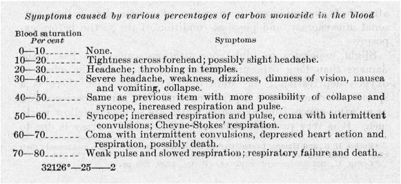
has discovered widely held beliefs regard- ing Carbon monoxide poisoning to be untrue during a 25-year career of research in the field. These include the mistaken beliefs that (1) symptoms correlate with presenting blood Carboxyhemoglobin levels, (2) residents are safe from CO poisoning if their home does not contain fuel-burning appliances, (3) Carboxyhemoglobin levels must be measured rapidly and on arterial blood, and (4) CO poisoning predisposes to premature long-term death from cardiac disease. In addition to providing the evidence disproving these myths, the importance of going back to the original reference when citing prior work is emphasized.

(C) 2015

Introduction

During a medical career, one will likely encounter “myths” from time to time. Myths can otherwise be described as facts that are widely ac- cepted as truisms despite a lack of supporting evidence to prove their validity. Myths are typically accepted as true because they are repeated- ly published in the literature or quoted by an authority in the field. When the naive individual questions the validity of a myth, instead of being provided supporting evidence, they are often told that “everyone” knows the fact to be true. It is thereby implied that if all others believe something, so should the individual.

In more than 25 years of research experience in the field of carbon monoxide poisoning, the author has recognized several “myths” in relation to the topic. In such instances, the goal should be to seek out the origin of the belief, disprove it when possible, help to discover the cor- rect facts, and publish them. This report describes some examples of the author’s personal experience in CO poisoning “myth busting.”

Some myths about CO poisoning

Myth 1: The carboxyhemoglobin level correlates with symptoms in Acute CO poisoning.

Articles and book chapters on CO poisoning have been published for decades containing tables or charts relating the degree of elevation of the Carboxyhemoglobin level to specific symptoms or signs in acute CO poisoning. These appear in both emergency medicine [1-3] and nonemergency literature [4,5]. Similar information is taught in

* Section of Pulmonary, Critical Care and Hyperbaric Medicine, Virginia Mason Medical Center, 1100 Ninth Ave, H4-CHM, Seattle, WA, 98101.

E-mail address: [email protected].

medical schools and is ubiquitous on the Internet. Interestingly, the ta- bles in the literature relating COHb levels to specific symptoms have common characteristics. First, most tables are remarkably similar, if not identical, in content, suggesting a common source. Second, a refer- ence for the information is typically not provided. When a citation is provided, it usually directs the reader to another paper containing the same table, this time without a reference.

A reference tree was constructed of 25 publications containing the usual table or a very similar one in an attempt to determine its origin [6]. All branches led back to publications containing the table but no cita- tion. A fortuitous discovery [7] revealed the same table published in a pamphlet by the US Bureau of Mines in 1923 (Figure) [8], again without a citation. However, when discussing absorption of CO by the blood, the report did refer to an earlier 1922 publication by the same authors enti- tled, “Physiological Effects of Exposure to Low Levels of Carbon Monox- ide” [9].

In their 1922 article, the investigators describe construction of a gas exposure chamber for humans. They then exposed themselves to CO con- centrations of 200-400 ppm, drew serial blood samples for COHb deter- mination, and recorded symptoms. Ten total exposures were conducted between the 3 authors, achieving peak COHb levels ranging from 16% to 28%. Symptoms recorded included “tightness across forehead,” “slight headache,” “dizziness,” and “throbbing headache,” among others. These were carried over to the COHb/symptoms table in their 1923 publication at respective COHb levels. It is unknown how they obtained the physio- logical responses to COHb levels from 30% to 80% reported in their table because no exposure resulted in COHb levels within that range.

Starting in August 2008, the Undersea and Hyperbaric Medical Soci- ety operated, in conjunction with the US Centers for Disease Control and Prevention, an online reporting system for prospective, real-time sur- veillance of CO-Poisoned patients treated with HBO2 [10]. In addition

http://dx.doi.org/10.1016/j.ajem.2015.10.051

0735-6757/(C) 2015

296 N.B. Hampson / American Journal of Emergency Medicine 34 (2016) 295297

Figure. Table of CO poisoning symptoms vs COHb levels from 1923 Bureau of Mines report [8].

to demographics, details of exposure, and laboratory results, symptoms were prospectively collected. Analysis of 2-year data from 1323 patients was performed to examine the relationship between COHb levels and symptoms [6]. The results showed no clear relationship between COHb and symptoms (Table), undoubtedly related to issues such as making the measurement at varying times postexposure and after dif- fering amounts of Oxygen administration. It is possible that peak COHb levels, if available, would correlate better with symptoms. Initial COHb levels upon ED presentation do not.

Myth 2: Without fuel-burning appliances in the home, there is no risk for CO poisoning.

Over the past several years, state legislation mandating residential CO alarms has virtually swept across the country. As of this writing in late 2015, 46 states have a law that requires a CO alarm in at least cate- gory of residence [11]. However, 33 (73%) of those exclude residences that do not have fuel-burning appliance, fireplaces, or attached garages from the requirement. This suggests that without a built-in source for CO generation, poisoning is not likely to occur. It ignores the possibility that a homeowner could unknowingly bring a source of CO into the home, such as a gasoline-powered electrical generator or charcoal grill. Although this places the responsibility for self-protection upon the resident of a detached, single-family home, such is not the case for mul- tifamily dwellings including duplexes, townhomes, or apartment build- ings. There have been numerous reports in the lay press where individuals located in several units of a multifamily dwelling have expe- rienced CO poisoning when a motor vehicle was left running in a single garage, a centrally located boiler malfunctioned, or a source of CO was brought into one residential unit. The usual explanation in these in- stances has been that CO traveled through connecting ductwork or common spaces such as stairways or hallways. However, sometimes, no pathway was apparent and CO seems to have traveled through walls. A study published in 2013 demonstrated that CO does have the poten- tial to pass through walls made of gypsum drywall, the most common type of wallboard used in the United States [12]. In this laboratory

investigation, it required only minutes for CO to diffuse across 0.25″ thick- ness, 0.5″ thickness, double-layer 0.5″ thickness, and painted double-layer 0.5″ thickness gypsum drywall. The reason for such permeability is that gypsum is porous, containing pores and channels that are approximately 1 million times the diameter of a CO molecule [12].

Because CO is able to diffuse through walls, individuals living in mul- tifamily dwellings cannot assume that they will be protected from CO poisoning because they do not have fuel-burning appliances or a fire- place within their residence. In reality, they may be dependent upon the activities of their neighbors to avoid CO exposure. Because of this and the possibility that a single-family homeowner may be unaware of the CO risk from an item brought indoors, CO alarms should be installed in all residences. You are still at risk for CO poisoning in your home even if fuel-burning appliances are not present.

Myth 3: Fresh arterial blood samples are needed for accurate determination of carboxyhemoglobin levels.

In some hospitals, the standard blood sample used for measurement of COHb levels is arterial [13]. Because of this, the sample is handled like an arterial blood gas sample, taken to the laboratory immediately and on ice. It is unclear how this practice evolved, but it is possible that be- cause levels of other gases in blood (eg, oxygen and carbon dioxide) are typically measured on arterial samples, someone assumed that CO should be also. This may be reinforced by the fact that many hospitals now have combination laboratory instruments containing both Blood gas analyzers and CO-oximeters. As such, both results are seen as com- ing from the same device.

Oxygen and carbon dioxide are measured on arterial blood because it reflects cardiopulmonary function better than venous blood. This is due to the fact that oxygen is consumed and carbon dioxide produced in peripheral tissues, changing the partial pressure of each as blood cir- culates from the arterial to Venous systems. Carbon monoxide is neither consumed nor produced to any significant degree in peripheral tissues. It has been demonstrated both in animal models and in humans that ar- terial and venous COHb levels are for clinical purposes identical [13,14].

Table

Prospectively collected symptoms of 1025 individuals poisoned with CO in comparison to presenting COHb level [6].

COHb

n

Headache

Dizziness

N/V

Confusion

Fatigue

Chest pain

SOB

LOC

0.0%-10.0%

98

65 (66%)

34 (35%)

49 (50%)

32 (33%)

39 (40%)

9 (9%)

9 (9%)

36 (37%)

10.1%-20.0%

313

181 (58%)

131 (42%)

130 (42%)

88 (28%)

79 (25%)

26 (8%)

22 (7%)

142 (45%)

20.1%-30.0%

368

243 (66%)

195 (53%)

158 (43%)

114 (31%)

114 (31%)

36 (10%)

38 (10%)

163 (44%)

30.1%-40.0%

183

85 (46%)

61 (33%)

60 (33%)

51 (28%)

25 (14%)

17 (5%)

19 (10%)

130 (71%)

40.1%-50.0%

63

17 (27%)

19 (30%)

15 (24%)

20 (32%)

15 (24%)

6 (10%)

5 (8%)

52 (82%)

N50.0%

10

1 (10%)

10 (100%)

1025

526

406

363

273

233

85

85

533

N/V = nausea/vomiting, SOB = shortness of breath, LOC = loss of consciousness.

N.B. Hampson / American Journal of Emergency Medicine 34 (2016) 295297 297

Rushing an iced blood sample to the laboratory is also not necessary. When anticoagulated blood samples with a COHb level approximately 30% were remeasured daily for a week and then weekly for a month, the COHb level did not change [15]. The result was not different wheth- er the samples were refrigerated or kept at Room temperature. The same study showed stability of COHb when anticoagulated samples were shipped without refrigeration from Seattle to Florida and back.

Measurement of COHb does not require fresh arterial blood. Carboxyhemoglobin can be measured in a delayed fashion on anticoagulated venous blood samples even if they have not been refrig- erated. Note that some laboratory CO-oximeter manufacturers recom- mend that the blood be anticoagulated with heparin (green top blood sample tube) [15].

Myth 4: CO poisoning predisposes to long-term risk for cardiac death.

This has been a common belief since the study by Henry et al was published a decade ago [16]. In that analysis of 230 consecutive CO- poisoned patients treated with Hyperbaric oxygen and hospitalized at a medical center in Minneapolis, mortality rates were compared be- tween those who experienced cardiac injury with their poisoning event and those who did not. It was demonstrated that “long-term” mortality was 38% in the myocardial injury group vs 15% in the noninjured group. When looking at the causes of deaths for those in whom it could be determined, 54% of those experiencing myocardial in- jury died of cardiac cause vs 27% in the noninjured group. This has been interpreted by some to demonstrate that CO poisoning predisposes to long-term risk of cardiac death.

However, Henry and coworkers did not separate short-term from long-term mortality [16]. Deaths occurring in hospital were included in the “long-term” mortality calculations. There were 6 deaths in hospi- tal in each group. Among those with myocardial injury, 4 were due to cardiac cause, as compared to 0 in the noninjury group. If these cases are considered short-term mortality and removed from posthospital survival analysis, those dying long term of cardiac cause in the myocar- dial injury group were similar to those in the noninjury group (50% vs 44%, respectively).

A subsequent study was performed at a regional treatment center for CO poisoning, analyzing data from 1073 patients treated over 28 years [17]. Deaths occurring within 90 days of poisoning were considered short term and were excluded. The analysis included 11,742 person- years of follow-up vs 1748 in Henry et al. Although an increased long- term mortality rate was seen in both accidentally and intentionally poi- soned individuals compared with the population, no evidence of excess cardiac mortality was seen among those with prior CO poisoning. Those

with unintentional poisoning had increased risk for long-term death from motor vehicle accidents, falls, and accidental poisoning, whereas those treated for intentional CO poisoning are at high risk for completed suicide. Neither group is at increased risk for cardiac mortality.

These are only a few of many myths about CO poisoning, and they serve to illustrate that myths likely exist in every discipline of medicine. The key to discovering and disproving myths is to go to the literature and find the original citation. It often does not exist or does not say what everyone believes. If you take the time to look for the original ev- idence, it is not difficult to become a myth buster.

References

  1. Flarity K. Environmental emergencies: carbon monoxide poisoning. In: Howard PK, Steinromm RA, editors. Sheehy’s emergency nursing: principles and practice. 6th ed. St. Louis: Moseby/Elsevier; 2010. p. 544-5.
  2. Olson KR. Carbon monoxide. In: Wolfson AB, editor. Clinical practice of emergency medicine. 4th ed. Philadelphia: Lippincott, Williams, and Wilkins; 1998. p. 1579.
  3. Varon J, Marik PE, Fromm RE, Gueler A. Carbon monoxide poisoning: a review for cli- nicians. J Emerg Med 1999;17(1):87-93.
  4. Devine SA, Kirkley SM, Palumbo CL, White RF. MRI and neuropsychological corre- lates of carbon monoxide exposure: a case report. Environ Health Perspect 2002; 110(10):1051-5.
  5. Consumer Product Safety Commission Staff.. Technology demonstration of a proto- type low carbon monoxide electrical generator. http://www.cpsc.gov/pagefiles/ 129846/portgen.pdf; 2012. [Accessed October 15, 2015].
  6. Hampson NB, Dunn SL, Members of the UHMS/CDC CO Poisoning Surveillance Group. Symptoms of acute carbon monoxide poisoning do not correlate with the initial carboxyhemoglobin level. Undersea Hyperb Med 2012;2(39):657-65.
  7. Donnay A. Carbon monoxide exposure and carboxyhemoglobin. Environ Health Perspect 2003;111:A511-2.
  8. Sayers RR, Yant WP. Dangers of and treatment for carbon monoxide poisoning. Bu- reau of Mines reports of investigations; 1923[serial number 2476, 11 pages].
  9. Sayers RR, Meriwether FW, Yant WP. Physiological effects of exposure to low levels of carbon monoxide. Public Health Rep 1922;37(19):1127-42.
  10. Hampson NB, Bell J, Clower JH, Dunn SL, Weaver LK. Partnering with a medical society to perform online disease surveillance. Undersea Hyperb Med 2012;39(2):647-55.
  11. Kidde, United Technologies. Home safety: fire safety laws and legislation. http:// www.kidde.com/home-safety/en/us/co-safety/carbon-monoxide-laws/. [Accessed September 3, 2015].
  12. Hampson NB, Courtney TG, Holm JR. Diffusion of carbon monoxide through gypsum drywall. JAMA 2013;310(7):745-6.
  13. Touger M, Gallagher EJ, Tyrell J. Relationship between venous and arterial carboxyhemoglobin levels in patients with suspected carbon monoxide poisoning. Ann Emerg Med 1995;25(4):481-3.
  14. Lopez DM, Weingarten-Arams JS, Singer LP, Conway EE. Relationship between arte- rial, mixed venous, and internal jugular carboxyhemoglobin concentrations at low, medium, and high concentrations in a piglet model of carbon monoxide toxicity. Crit Care Med 2000;28:1998-2001.
  15. Hampson NB. Stability of carboxyhemoglobin in stored and mailed blood samples. Am J Emerg Med 2008;26:191-5.
  16. Henry CR, Satran D, Lindgren B, Adkinson C, Nicholson TI, Henry TD. Myocardial in- jury and long-term mortality in moderate to severe carbon monoxide poisoning. JAMA 2006;295:398-402.
  17. Hampson NB, Hauff NM, Rudd RA. Increased long-term mortality among survivors of acute carbon monoxide poisoning. Crit Care Med 2009;37(6):1941-7.